

Fluorescent labelled E. coli can be seen in the swollen pharynx of some dying C. elegans (P, left) but not others with an atrophied pharynx (p, right).
With improving healthcare humans are living longer than ever before, but with longer life comes ever more senescence-related pathologies. Understanding the role genes and environment play in the development of such pathologies in the complex system of our bodies is difficult. The nematode Caenorhabditis elegans is a great model organism and has been used extensively to study the biology of ageing because just like more complex animals, C. elegans also develop senescent pathologies.
Researchers from the University College London and Washington University, MO studied age-related pathologies in C. elegans and their role in limiting lifespan. Understanding these processes in a simple organism may in turn help to further understand the origins of human age-related pathologies.
To investigate the causes of death, the group analysed the corpses of recently expired wildtype C. elegans and found two particular forms with different pharyngeal pathologies. One type, named P ("big P") death, occurred earlier than the other and had increases in the posterior bulb size of 20-120%. The other, named p ("small p") death, showed a shrinking of the posterior bulb by up to 70%.
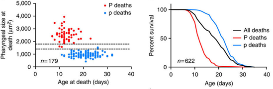
The figures above show the age distribution and percentage survival of P and p deaths.
Dissections and RFP labelling of the E. coli food source established that the enlarged posterior bulb in P death individuals was due to E. coli infection in elderly C. elegans. The high pharyngeal pumping rates typical of young nematodes is thought to mechanically damage the cuticle, creating vulnerability to invasion. Their findings suggest there is a narrow time frame in which young nematodes are thus susceptible. Consistent with this, the group found that mutants with reduced pumping rate had fewer P deaths, and lived longer. However, it was also found that worms dying with P and p death previously had similar pumping rates, so why did some worms but not others get an infected pharynx?
The difference appeared to be due to the ability of some nematodes (p death type) to heal the cuticle thereby preventing invasion.
When asked about their work and the role of the PE120 stage, Professor David Gems stated, “to help identify old age pathologies that limit life, we watched nematodes as they aged, measuring a range of pathologies, and then measured their lifespan. By measuring how well each pathology correlated with lifespan we could identify pathologies likely to cause death. But to do this required repeatedly putting immobilized nematodes under the microscope, and we needed to do this in a way that wasn’t so stressful that it shortened their lifespan. By using the PE120 Linkam stage to gently cool the worms, we were able to avoid using stressful anaesthetics. We were able to confirm that repeated viewing of nematodes using the PE120 Linkam stage in this way did not shorten their lifespan.”
The group used a novel approach to understand ageing by analysing and combining pathology and mortality profiles. Further work can now be conducted with a view to understanding how genes that affect lifespan differentially affect worms dying from different causes.
By Tabassum Mujtaba


