Exploring plasmon-actuated nano-asssembled microshells using a Linkam hotstage

LTS420 shown above

Nanomaterials have been found to have interesting electronic, magnetic and optical properties. They can manipulate electromagnetic fields through localised surface plasmon resonance to modulate light interactions. Such plasmonic phenomena are popular in application for the biomedical field. 

Researchers from the University of California, Merced and Stanford University in the USA developed a micro-scale delivery module for various organic and inorganic compounds using nanomaterials. 

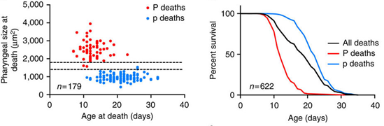
Their aim was to create something that would be versatile and capable of encapsulating a range of different materials (drugs, dyes, cells, bacteria, etc.) for many different applications. These could include drug delivery for cancer treatment, releasing dyes in vivo for fluorescence imaging, or tissue engineering. The problem with existing platforms is that they are either leaky, unable to hold the contents without loss for any prolonged period, or they are incapable of releasing contents in a spatially and temporally controlled manner. For example, other cargo delivery systems that use light to activate the release of the cargo need several milliwatts of power over several minutes to achieve the required effect, therefore creating significant localised heating. The group managed to reduce the power required to less than 2 mW and the release time to under 5 seconds. As a result, the total temperature increase at the vicinity of the capsules is only to ~ 40°C, which is well within tolerable limits for many biological systems.

The team used an LTS350*,(now superseeded by the LTS420), for their experiments. When asked on the importance of the hotstage, Dr Ghosh said: “One of the most critical parameters that determine whether a cargo delivery system is viable in vivo is the thermal gradient that is produced because of the photothermal effect when optical excitation used to rupture the shells is in resonance with the plasmonic response of the nanoparticles that make up the shell walls. To estimate this, the first step was to use heat to rupture the shells instead of light. That is where we used the heating stage.”
 

Fluorescence microscopy images of a Nano-Assembled Microshell loaded with a fuorescent dye on the LTS350 stage. 

Fluorescence microscopy images of a Nano-Assembled Microshell loaded with a fuorescent dye on the LTS350 stage. 

Their method has proved to be exciting and advantageous. No leakage was seen for over five months after encapsulation, promising a long shelf life. Furthermore, a lower optical intensity was required for shell disintegration compared to other methods. 

Although more work is required to improve future in-vivo applications (such as actuation by near infra-red and reducing overall size of capsule), their work is a promising result for future cargo delivery systems. 

Find out more about the research here

*The LTS350 has been superseded by the LTS420 offering a large temperature range and better temperature control to 0.01°C.
 

Studying age-associated pathologies through temperature controlled microscopy

Fluorescent labelled E. coli can be seen in the swollen pharynx of some dying C. elegans (P, left) but not others with an atrophied pharynx (p, right).

Fluorescent labelled E. coli can be seen in the swollen pharynx of some dying C. elegans (P, left) but not others with an atrophied pharynx (p, right).

With improving healthcare humans are living longer than ever before, but with longer life comes ever more senescence-related pathologies. Understanding the role genes and environment play in the development of such pathologies in the complex system of our bodies is difficult.  The nematode Caenorhabditis elegans is a great model organism and has been used extensively to study the biology of ageing because just like more complex animals, C. elegans also develop senescent pathologies.

Researchers from the University College London and Washington University, MO studied age-related pathologies in C. elegans and their role in limiting lifespan. Understanding these processes in a simple organism may in turn help to further understand the origins of human age-related pathologies.

To investigate the causes of death, the group analysed the corpses of recently expired wildtype C. elegans and found two particular forms with different pharyngeal pathologies. One type, named P ("big P") death, occurred earlier than the other and had increases in the posterior bulb size of 20-120%. The other, named p ("small p") death, showed a shrinking of the posterior bulb by up to 70%.

The figures above show the age distribution and percentage survival of P and p deaths.

The figures above show the age distribution and percentage survival of P and p deaths.

Dissections and RFP labelling of the E. coli food source established that the enlarged posterior bulb in P death individuals was due to E. coli infection in elderly C. elegans. The high pharyngeal pumping rates typical of young nematodes is thought to mechanically damage the cuticle, creating vulnerability to invasion. Their findings suggest there is a narrow time frame in which young nematodes are thus susceptible. Consistent with this, the group found that mutants with reduced pumping rate had fewer P deaths, and lived longer.  However, it was also found that worms dying with P and p death previously had similar pumping rates, so why did some worms but not others get an infected pharynx? 

The difference appeared to be due to the ability of some nematodes (p death type) to heal the cuticle thereby preventing invasion. 

When asked about their work and the role of the PE120 stage, Professor David Gems stated, “to help identify old age pathologies that limit life, we watched nematodes as they aged, measuring a range of pathologies, and then measured their lifespan. By measuring how well each pathology correlated with lifespan we could identify pathologies likely to cause death. But to do this required repeatedly putting immobilized nematodes under the microscope, and we needed to do this in a way that wasn’t so stressful that it shortened their lifespan. By using the PE120 Linkam stage to gently cool the worms, we were able to avoid using stressful anaesthetics. We were able to confirm that repeated viewing of nematodes using the PE120 Linkam stage in this way did not shorten their lifespan.”

The group used a novel approach to understand ageing by analysing and combining pathology and mortality profiles. Further work can now be conducted with a view to understanding how genes that affect lifespan differentially affect worms dying from different causes.

By Tabassum Mujtaba

Zhao, Y. et al. Two forms of death in ageing Caenorhabditis elegans. Nat. Commun. 8, 15458 doi: 10.1038/ncomms15458 (2017).

Investigating meniscus-assisted solution printing of large-grained perovskite film for high-efficiency solar cells

LTS420

Solution printing is a novel technique which uses an ink solution, containing semi-conductor precursors or nanoparticles, and deposits these on substrates with desirable characteristics. This offers a cost-effective method of creating large area thin film optoelectronics whilst also offering precise control over the stoichiometry and adaptability of the material. Metal halide perovskites have superb optoelectronic properties and the last few years has seen their power conversion efficiency increase rapidly, largely through the optimisation of the crystal morphology. 

The requirement to control morphology has posed a problem for solution printing. The understanding of crystallisation in dynamic flow of perovskite inks is quite limited, thus imposing restrictions in achieving high-quality perovskite films by the solution-printing technique.

A group from the Georgia Institute of Technology and the University of Nebraska-Lincoln in the USA used a meniscus-assisted solution printing method to elucidate the crystallisation kinetics of perovskite inks and help create high efficiency perovskite solar cells. The thin-films were created with preferred crystal orientation with micrometre-scale grains. 

When discussing their work Professor Lin said, “Through integrating the meniscus effect within the solution printing, we found that the solvent evaporation could be largely promoted by the meniscus effect instead of the thermal evaporation as in conventional doctor-blade coatings, thus leading to the low-temperature solution-based deposition of high-quality perovskite films with preferred crystal orientations. This low temperature feature circumvents thermal degradations and thermomechanical fatigues on perovskite and electrode materials, as well as decreases energy consumptions. Our technique paves the pathway for depositing perovskite thin films on flexible polymer substrates, and is anticipated to promote the future development and applications of perovskites in low-cost, large-area, and flexible optoelectronic devices.”

The group used an LTS350*, now superseded by the LTS420 to control the substrate temperature during the meniscus-assisted solution printing process, due to its capability of precisely controlling the temperature at ±0.1oC. Their investigation on the crystallisation kinetics of perovskite films revealed that a large temperature fluctuation would seriously impact the crystallisation kinetics of perovskite films during the meniscus-assisted printing process. The LTS350* was ideal for maintaining the substrate at a constant temperature and focusing on the exploration of the meniscus effect on the perovskite crystallisation process. 

Their study helped to uncover the crystallisation kinetics of perovskites during the printing process, providing rational guides to precisely control the crystallisation morphology of printed perovskite films. By improving the control over morphology, the group’s work helps to pave a route to large-area optoelectronic devices for commercial applications.

*The LTS350 has been superseded by the LTS420 offering a large temperature range and better temperature control to 0.01°C.

Read the research paper here

Investigating the Anaerobic Consortia of Fungi and Sulfate-reducing Bacteria in Deep Granite Fractures using Linkam’s THMS600.

The deep biosphere is one of the least understood ecosystems, despite being considered vital for energy cycling of the earth. It is thought to have approximately 19% of the earth’s biomass yet samples are hard to come by, making their study difficult. Microorganisms from the deep biosphere that have been studied are generally prokaryotes, with microeukaryotes being largely ignored.

Recently samples were taken from a 740m deep drill core sample in Sweden after the site was investigated for its suitability for deep nuclear waste repositories. Findings have shown the presence of fossil and active fungi in these deep ecosystems, but little work has gone into understanding them.

Drake et al., studied the microorganisms in these deep crystalline fractured rock samples. Their aim was to gain a better understanding of the microbial processes in the continental crust. The knowledge of this vast realm is very scarce and tells us more about life forms and processes under extreme conditions which may also have important implications for nuclear waste storage. 

Their analyses found the microorganisms belonged to the Kingdom Fungi and were found to be anaerobic. The closest systems studied were that of anaerobic fungi in the rumina of ruminant animals. It was proposed that the fossilised fungi also shared a symbiotic relationship with bacteria in the deep biosphere. 

The group used a THMS600 to help indicate the approximate age of the fungi. Dr Drake said, “The THM600 was used to investigate fluid inclusions in calcite crystals that were spatially related to the fungi. The fluid inclusion signatures gave us information about past conditions (e.g. salinity) in the fracture void. Because no radiometric dating could be made of the fungi, the fluid inclusion signatures (when put in a paleohydrogeological context) serve as an important temporal indicator for when the fungi were active.” 

Their work highlighted an intimate relationship between the fungi and sulphate reducing bacteria, further drawing attention to the richness of the deep oligotrophic biosphere which is often neglected. These fungi were found to provide significant amounts of H2 to autotrophic microorganisms in the crystalline continental crust.

The group also looked at the biochemistry of these fungi and found they may pose a threat to repositories of toxic waste.  This is through either directly breaking down the barriers holding the waste, or by facilitating the bacterial community into doing so. 

Their work highlights the importance of studying these neglected geological microorganisms. With fossil fuels running out, nuclear energy may be the way forward. But to safely store away waste products, understanding their chemical and geological environment is of utmost importance as illustrated by Drake et al., As such it becomes vital to study ecosystems, such as the deep biosphere, in its entirety. 

Read the research paper here